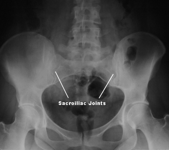
Sacroiliac SI joints and the first two segments of sacrum must be in center of radiograph or in the collimated field. -shoulder thorax and thigh supported foam -saggital plane 1 medial to the ASIS of affected side centered to grid.

10 x 12 film 2.
Si joints x ray positioning. It is also used to determine any dislocation or subluxation to the joint. Patient positioned supine on the imaging table with legs extended. Elevate the side of interest approximately 25 to 30 2.
The patients body should be adjusted to allow the bodys long axis to be parallel to the long axis of the x-ray table. SACROILIAC JOINTS SI JOINTS ROUTINE VIEWS. 10 x 12 film 2.
15o cephalic angle enter halfway between ASIS. Obliques RPO and LPO 1. 10 x 12 film 2.
Patient rotated 30o from AP position 4. Correct Central Ray angulation when sacroiliac joints spaces and the L5 to S1 junction and sacral foramina should appear open. Sacroiliac SI joints and the first two segments of sacrum must be in center of radiograph or in the collimated field.
A cassette with an X-ray film is placed where the upper posterior protrusions awns are projected on the crest of the iliac spine and the X-ray machine beam is focused from a meter distance to the abdominal cavity - slightly to the side of its midline at the level of the upper anterior os ilium awns. The central-ray angle for an AP oblique sacroiliac joint is. How many true or movable vertebrae are there in the vertebral column.
If SI joints are imaged in the PA position describe the central ray. Perpendicular for obliques 35 degrees caudal if PA. Review of Radiographic Anatomy Positioning and Pediatric Positioning.
Jeana Fleitz MEd RTRM The X-Ray Lady. Clarks Positioning in Radiography 12th ed Arnold PDF Clarks Positioning in Radiography 12th ed Arnold indah apriyani - Academiaedu Academiaedu no longer supports Internet Explorer. The sacroiliac joint is composed of three portions oriented in different planes.
To study the sacroiliac joints it seems desirable to obtain an anteroposterior view of the pelvis with the patient in a lithotomy position. Then if needed each joint can be radiographed separately by using oblique views. The iRadTech app is a radiographic positioning guide for Apple and Android smart phones and tablets.
It is also available as a web app delivered to a browser which is platform independent. More than 220 radiographic positions described with detailed instructions and human model and x-ray images. Position of part AP oblique RPOLPO SI joint.
-Side being examined is farthest from table. -MCP 25-30 to table. -shoulder thorax and thigh supported foam -saggital plane 1 medial to the ASIS of affected side centered to grid.
-Check for rotation along back. Other and on phantoms in energized X-ray labs. This class must be taken concurrently with RAD 112L.
If RAD 112L is dropped then RAD 112 must be dropped. This course requires student to student or instructor to student physical contact appropriate and necessary to demonstrate teach or learn the principles of radiographic positioning. What is Sacroiliac Joints X-Ray.
Sacroiliac Joints or SIJ X-Ray may be performed to evaluate the cause of pelvic pain. It may be used to detect foreign objects stones and the cause of pain and swelling. Your SI joints are located where the sacrum and ilium meet.
Anatomy and physiology of the sacroiliac joints. The sacroiliac joint is a true diarthrodial joint that joins the sacrum to the pelvis. The sacrum tailbone connects on the right and left sides of the ilia pelvic bones to form the sacroiliac joints.
The pelvic girdle is made up of two innominate bones the iliac bones and the sacrum. DIGITAL X-RAY ARTICLES Proper positioning for the pelvis and proximal femur The lowdown on lumbar spine positioning Radiographic positioning techniques for the cervical spine Boning up on humerus clavicle and AC joint positioning Getting the most from shoulder positioning The bends and flexures of forearm and elbow x-ray positioning. Position of patient Supine or prone.
Injured patients should NOT be turned over. In trauma patients a lumbar spine X-ray is done in the AP or PA position with minimal movement of the patient. Position of part The patients knees are bent to ensure the back is flat on the table.
The sacroiliac joints are seen at an angle on the AP radio-graph resulting in some overlap of structures. The normal sacroiliac joint is a syndesmotic joint in its upper two-third and a synovial joint in its lower anterior one-third5 The lower sacroiliac joint is well seen on a pelvic outlet view2 A. The thumb is imaged in two directions.
For the anterior-posterior AP image the arm is rotated internally with the dorsal side of the thumb flat on the x-ray plate. The x-ray beam will pass through the hand from palmar to dorsal fig. For the lateral image of the thumb the other fingers are abducted to ulnar and an attempt is made to.
Central Ray CR perpendicular to IR directed to PIP joint Minimum SID of 40 inches 100cm Collimation. Collimate on four sides to affected finger. Second Digit Positioning For second digit a mediolateral is advised if the patient can assumed this position.
Place the second digit in contact with cassette. Definition of image is improved with less OID. The diagnosisclassification of ankylosing spondylitis typically depends on key features on the X-ray.
These features being sclerosis thickening of the adjacent bone erosion irregularity of the bone and joint space narrowing. Show the X-ray to a bunch of radiologists without priming them with a focused history and you are very likely to.