It is commonly used in the practical labs to study organisms behaviour and characteristics like size shape and arrangement. The lens system responsible for enlarging the image.

The lighting system that provides the light source and its regulation.
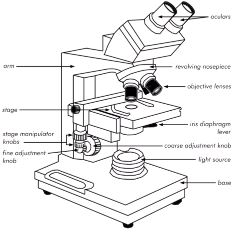
Bright field microscope parts. A bright-field microscope is consists of A piece of apparatus consisting of an eyepiece an objective lens a condenser lens stage and light source which collects electromagnetic radiation in. Figure 31 Parts of a brightfield microscope. Base - The part of the microscope that rests on the table is called the base.
Light Source - The illuminator for your microscope is built into the base and is controlled by an onoff switch. Arm - The arm is a vertical support that connects the base of. Parts of Brightfield Microscope Compound Light Microscope Bright field Microscope Components.
The bright field microscope is composed of several components including. Eyepiece Ocular lens The microscope contains two eyepiece lenses at the top which concentrate the image from the objective lenses. With your eyes this is where you view the formed image.
The brightfield microscope is made up of various parts including Eyepiece Ocular lens it has two eyepiece lenses at the top of the microscope which focuses the image from the objective lenses. This is where you see the formed image from with your eyes. As brightfield is the most widely used type of microscopy other types of microscopes can be adapted to brightfield.
The microscope consists of three well-defined parts. The lens system responsible for enlarging the image. The lighting system that provides the light source and its regulation.
Eye piece used to view the magnified image from the slide. Holds various objective lenses and rotates to magnification. Allows to magnify the specimen.
Brightfield Light Microscope Compound light microscope This is the most basic optical Microscope used in microbiology laboratories which produces a dark image against a bright background. Made up of two lenses it is widely used to view plant and animal cell organelles including some parasites such as Paramecium after staining with basic stains. Microscope Parts Drawing Free Download On Clipartmag.
Nikon Optiphot Compound Brightfield Microscope Laboratory. Olympus Bh2 Research Microscope Cutaway Diagram Olympus Life Science. Science Cartoon Clipart Science Light Drawing Transparent.
User Manual Zeiss Axiostar Microscope. 9 Eye PieceThe part you look at with your eye. Usually 10 X magnification.
Click Here to Return to the Main Slide Click Here to Return to the Main Slide 10 Arm Used to safely transport microscope. Start studying Parts of the Bright Field Microscope. Learn vocabulary terms and more with flashcards games and other study tools.
What are the 3 Basic Parts of a Microscope. The main parts of a microscope are easy to identify. The upper part of the microscope that houses the optical elements of the unit.
The bottom of the microscopewhat the microscope stands on. Structural element that connects the head of the microscope to the base. Other Important Parts and Their Functions.
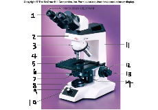
Parts of Microscope. 1 Eyepiece 2 Nose piece 3 Objective lens 4 Stage clips or Slide holder 5 Aperture 6 Stage 7 Adjustment knobs 8 Stage controls 9 Condenser lens 10 illuminator The Eyepiece Lens. It also termed ocular located at the top of the microscope.
What are the parts of a brightfield microscope. Brightfield microscopes are made up of several different parts and what these depend on what type of brightfield microscope you are looking at. That said the most essential parts are the light source and the lenses.
Bright-field microscope is a compound light microscope which illuminates the background against a stained specimen. It is commonly used in the practical labs to study organisms behaviour and characteristics like size shape and arrangement. It is a type of light microscopy where a path of light is very simple which requires a light source like a halogen lamp condenser lens objective lens and ocular lens.
The compound microscope is also known as the bright-field microscope because the light passes directly through the light source to the eye through the two lenses. This mechanism makes the field of vision brightly illuminated. Parts of Compound Microscope.
OBSERVED USING BRIGHT-FIELD MICROSCOPY AND APPROPRIATE MAGNIFICATIONS Prepared slides stained - bacteria 1000x thick tissue sections 100x 400x thin sections with condensed chromosomes or specially stained organelles 1000x large protists or metazoans 100x. Smears stained - blood 400x 1000x negative stained bacteria 400x 1000x. Handling a Microscope Always carry a microscope with one hand on the arm and the other under the base.
Always use lens paper to clean the objectives and eye pieces of a microscope. Neveruse paper towels Kimwipes etc. Before storing the microscope always clean it.
The light path of a bright-field microscope is extremely simple no additional components are required beyond the normal light-microscope setup. The light path therefore consists of. A transillumination light source commonly a halogen lamp in the microscope stand.
A condenser lens which focuses light from the light source onto the sample. Microscope Parts III Pull the camera slider arrow all the way out before you take a picture to direct the light to the cameras. Turn the black knob red circle to select the camera you want to use.
Black white camera. Formation in the light microscope. Types of Specimens for Bright Field Microscopy The best optics and the best instrument alignment are useless if there is no visual difference between the specimen and its surroundings or among the various parts of the specimen.
Human vision is sensitive to differences in brightness amplitude and color. The Bright-field Compound Mieroscope. Using the following list label the parts of the bright-field compound microscope.
Base dialirheostat iris diaphragm light souree mechanical stage knobs objeetive ocular pinion knob revolving nosepiece stage body tube condenser lens coarse adjustment knob fine adjustment knob illuminator 2.